Reduced Dose Load
MammoExpress implements a patented narrow beam X-ray scanning technology, which effectively eliminates scattered radiation, which leads to a reduction in radiation exposure and improves overall image contrast. In most mammography systems, scattered radiation increases the noise level in the image, which requires compensation by adding a screening grid.
Reliable Scanning
MammoExpress tumor scanner uses the TDI (“delayed accumulation”) operating principle, which eliminates non-functional pixels and provides excellent image quality at a minimum dose. During scanning, each object appears more than a hundred times, thus providing a representation of the image.
Automated Examination Process
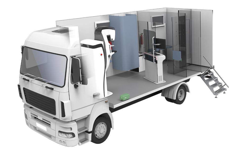
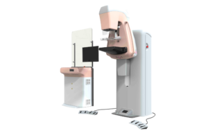
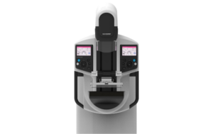
Isocentric rotation motorized movements combined with customizable presets of the inspection and diagnostic process significantly reduce examination time. Positioning control panels, accessible from all sides of the device, including the top of the device, make lateral positioning more convenient. The automatic decompression function releases the patient’s chest immediately after exposure for added comfort.
Tungsten Anode
Image quality and the patient’s radiation dose are reacting factors in mammography. MammoExpress uses an X-ray tube with a tungsten anode, which reduces the patient’s radiation exposure by using the optimal radiation spectrum.
Award-winning Software
The patient-centric design of the MammoExpress has been recognized and features such as side handles help the patient maintain stability during both cranio-sacral (SS) and medial lateral (MLO) projection. The curved surface of the detector increases the comfort level for the patient during the examination.
Fully Integrated Solution
MammoExpress can be supplied as a complete diagnostic system, including a diagnostic workstation, a PACS server, a NAS storage device, a dry-print medical printer and CAD software. With this configuration, MAMMOSCAN can be used in any clinic or radiology center as a stand-alone diagnostic device with full capabilities.
The acquisition and analysis of radiographic images is managed by software from automated workstations.